Image for Molecular imaging in oncology

Molecular imaging in oncology - 187

Jackson, Edward F.Kim, E. EdmundAoki, J.(Contributions by)Baghaei, H.(Contributions by)Ilgan, S.(Contributions by)Inoue, T.(Contributions by)Li, H.(Contributions by)Uribe, J.(Contributions by)Wong, F.C.L.(Contributions by)Wong, W.-H.(Contributions by)Yang, D.J.(Contributions by)
Part of the Recent Results in Cancer Research series
See all formats and editions

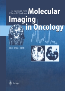
Advanced routine imaging techniques have made it possible to diagnose localized abnormalities often before they have produced irreversible damage.

PET permits visualization of the metabolic or physiologic characteristics of tumors.

MRI shows exclusive morphologic abnormalities, and functional MRI depicts the motor and sensory status in the vicinity of tumors.

MRS offers biochemical information of tumors and surrounding normal tissues.

Combining PET, MRI and MRS information we should be able to differentiate tumors from non-tumor lesions, to characterize types or grades of tumors, and to monitor tumor regression, recurrence or response to therapy.

This book reports updated techniques, instrumentation and clinical application of PET, MRI and MRS in cancer management.

Read More
Special order line: only available to educational & business accounts. Sign In
£89.50
Product Details
Springer
364259879X / 9783642598791
eBook (Adobe Pdf)
06/12/2012
Germany
English
1 pages
Copy: 10%; print: 10%
Description based on CIP data; resource not viewed.